| Specificity |
The antibody is specific for proteins containing phosphotyrosine residues and phosphotyrosine peptides. It does not cross-react with phosphoserine, phosphothreonine or nonphosphorylated tyrosine residues. |
| Host Species |
Mouse |
| Immunogen |
Phosphotyrosine containing carrier protein |
| Conjugate |
Unconjugated |
|
Working concentrations for specific applications should be determined by the investigator. The appropriate concentrations may be affected by secondary antibody affinity, antigen concentration, the sensitivity of the method of detection, temperature, the length of the incubations, and other factors. The suitability of this antibody for applications other than those listed below has not been determined. The following concentration ranges are recommended starting points for this product. |
| Application |
Recommended Usage |
| Western Blot |
1-2 μg/ml |
| Immunoprecipitation (IP) |
1-4 μg/ml |
| Immunocytochemistry/Immunofluorescence (ICC/IF) |
4 μg/ml |
| Flow Cytometry |
4 μg/ml |
| ELISA |
0.1 μg/ml |
|
| Form |
Lyophilized |
| Storage Buffer |
lyophilized with PBS, pH 7.4, containing 0.02% sodium azide.Note GenScript can customize this product per customer's request including product size, buffer components, etc. |
| Reconstitution |
Reconstitute the lyophilized powder with deionized water (or equivalent) to an final concentration of 0.5 mg/mL. |
| Storage Instructions |
The lyophilized product remains stable up to 2 years at -20°C from date of receipt. Upon reconstitution, it can be stored for 2-3 weeks at 2-8°C or for up to 12 months at -20°C or below. Avoid repeated freeze and thaw cycles. |
| Purification |
Protein A affinity column |
| Isotype |
Mouse IgG1, κ& IgG2a,κ |
| Clonality |
Cocktail |
| Clone ID |
Not applicable |
plus")
Western blot analysis of tyrosine phosphorylation status in different cells with THETM Phosphotyrosine Antibody (E10)plus (A01819, 1 μg/ml)
1. EGF-stimulated A431 cell lysates (50 μg).
2. Untreated A431 cell lysates (50 μg).
3. Insulin-stimulated HEK293 cell lysates (50 μg).
4. Untreated HEK293 cell lysates (50 μg).
The signal was developed with IRDyeTM800 Conjugated affinity Purified Goat Anti-Mouse IgG.
plus")
Western blot comparison of a panel of phosphotyrosine antibodies with EGF-stimulated A431 cell lysates. THETM Phosphotyrosine Antibody (E10)plus detects the most bands representative of various phosphotyrosine residues.
1. Phosphotyrosine Antibody (5E10), mAb, Mouse (A01817, 1 μg/ml)
2. Phosphotyrosine Antibody(18E10), mAb, Mouse (A01818, 1 μg/ml)
3. THETM Phosphotyrosine Antibody (E10)plus (A01819, 1 μg/ml)
4. Anti-Phosphotyrosine, 4G10 ? Platinum (Millipore, 05-1050, 1:1000)
The signal was developed with IRDyeTM800 Conjugated affinity Purified Goat Anti-Mouse IgG.
plus")
Flow cytometric analysis of EGF-stimulated A431 cell and untreated A431 cell with THETM Phosphotyrosine Antibody (E10)plus (A01819, 4 μg/ml) (red and black respectively).
The signal was developed with FITC conjugated Goat Anti-Mouse IgG.
plus")
ELISA analysis of various phosphotyrosine peptides and corresponding non-phosphorylated peptides with THETM Phosphotyrosine Antibody (E10)plus (A01819, 0.1 μg/ml). Results demonstrate the antibody has excellent recognition of different phosphotyrosine peptides and no cross-reactivity with non-phosphorylated peptides.
plus")
Immunocytochemistry/Immunofluorescence analysis of EGF-stimulated A431 cell and untreated A431 cell with THETM Phosphotyrosine Antibody (E10)plus (A01819, 4 μg/ml).
The signal was developed with FITC conjugated Goat Anti-Mouse IgG.
| Target Background |
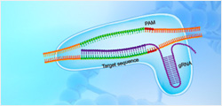
Phosphotyrosine is a tyrosine residue covalently bound to a phosphate via its hydroxyl group. A tyrosine kinase catalyzes the transfer of a phosphate from ATP to a tyrosine residue on the protein substrate. Tyrosine phosphorylation plays a key role in intracellular signaling and cancer development. Many tyrosine kinases are drug targets for different forms of cancer. Phosphotyrosine antibody specifically binds to phosphotyrosine residues. It is a valuablel tool to analyze tyrosine phosphorylation and monitor the activity of tyrosine kinase in high throughput drug discovery. THE? Phosphotyrosine Antibody (E10) plus recognizes a broadspectrum of phosphotyrosine-containing proteins from numerous species.GenScript THE? Phosphotyrosine Antibody (E10) plus is an anti-Phosphotyrosine monoclonal antibody (5E10E1) mixed with another anti-Phosphotyrosine monoclonal antibody (18E10D2) at an optimal ratio. The antibodies are produced from a hybridoma resulting from the fusion of SP2/0-Ag14 myeloma and B-lymphocytes harvested from mouse immunized with phosphotyrosine conjugated to KLH. |
| Synonyms |
Mouse monoclonal to Phosphotyrosine/pY |
For laboratory research use only. Direct human use, including taking orally and injection and clinical use are forbidden.